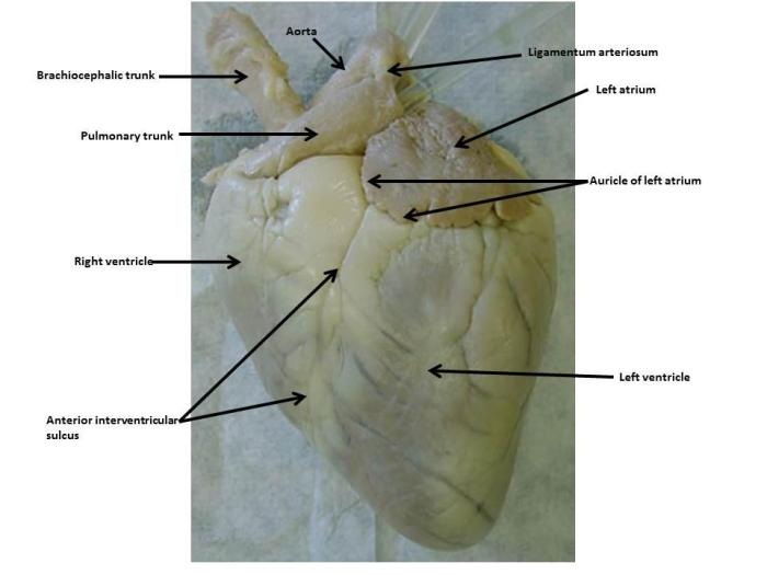
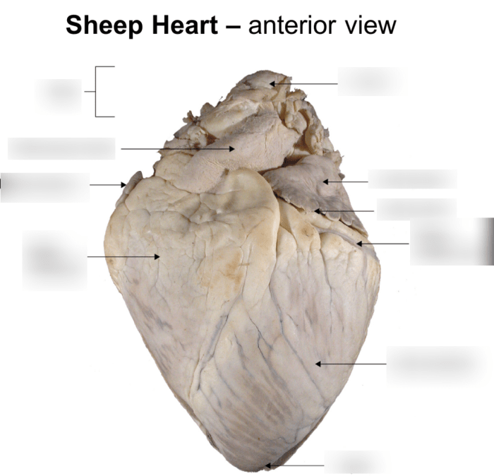
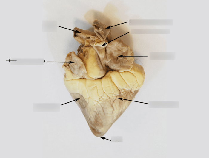
Anterior view of sheep heart – The anterior view of the sheep heart offers a captivating glimpse into the intricate workings of this vital organ. As we delve into its anatomy, we’ll explore the structures that orchestrate the heart’s rhythmic beat and the vessels that sustain its lifeblood.
The heart’s anterior surface is dominated by the right and left atria, responsible for receiving blood from the body and lungs, respectively. Below them lie the right and left ventricles, the muscular chambers that pump blood throughout the circulatory system.
Anterior Surface of the Sheep Heart
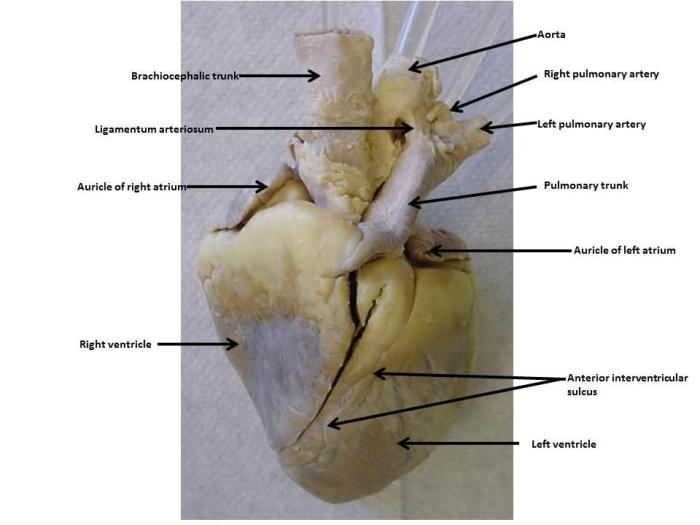
The anterior surface of the sheep heart is a complex structure that houses several crucial components responsible for the heart’s pumping action. These structures include the right atrium, right ventricle, left atrium, and left ventricle, each playing a distinct role in the heart’s overall function.
Right Atrium
- Located in the upper right portion of the heart.
- Receives deoxygenated blood from the body via the superior and inferior vena cavae.
- Acts as a temporary reservoir for blood before it enters the right ventricle.
Right Ventricle
- Situated below the right atrium.
- Pumps deoxygenated blood to the lungs via the pulmonary artery for oxygenation.
- Contains the tricuspid valve, which prevents backflow of blood into the right atrium.
Left Atrium
- Positioned in the upper left portion of the heart.
- Receives oxygenated blood from the lungs via the pulmonary veins.
- Acts as a temporary storage for blood before it enters the left ventricle.
Left Ventricle
- Located below the left atrium.
- Pumps oxygenated blood to the body via the aorta.
- Contains the mitral valve, which prevents backflow of blood into the left atrium.
Cardiac Vessels and Nerves
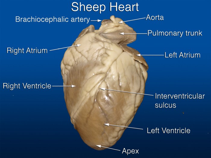
The anterior surface of the sheep heart is richly supplied by a network of blood vessels and nerves. These structures play a vital role in ensuring the proper functioning of the heart muscle, supplying it with oxygen and nutrients, and regulating its activity.
Coronary Arteries
The coronary arteries are the major blood vessels that supply oxygenated blood to the heart muscle. The two main coronary arteries are the left and right coronary arteries, which arise from the aorta just above the aortic valve. The left coronary artery supplies blood to the left atrium, left ventricle, and the anterior surface of the right ventricle.
Before you dive into the aphg unit 5 practice test , let’s take a quick look at the anterior view of a sheep heart. Notice the atrioventricular groove and the coronary sulcus. These features are crucial for understanding the heart’s anatomy and function.
The right coronary artery supplies blood to the right atrium, right ventricle, and the posterior surface of the left ventricle.
Coronary Veins, Anterior view of sheep heart
The coronary veins collect deoxygenated blood from the heart muscle and return it to the right atrium. The major coronary veins are the great cardiac vein, the middle cardiac vein, and the small cardiac vein. The great cardiac vein drains the anterior surface of the heart, while the middle cardiac vein drains the posterior surface of the left ventricle and the small cardiac vein drains the right ventricle.
Innervation of the Heart
The anterior surface of the heart is innervated by the vagus nerve and the sympathetic nerves. The vagus nerve is part of the parasympathetic nervous system and it slows down the heart rate and decreases the force of contraction. The sympathetic nerves are part of the sympathetic nervous system and they increase the heart rate and increase the force of contraction.The
innervation of the heart is essential for regulating heart function. It allows the heart to respond to changes in the body’s needs, such as during exercise or when at rest.
Pericardial Sac
The pericardial sac is a double-walled membrane that envelops the heart and its major blood vessels. It consists of an outer fibrous layer, the fibrous pericardium, and an inner serosal layer, the serous pericardium.
The pericardial sac provides several important functions:
- Protection:The fibrous pericardium protects the heart from external trauma and mechanical injury. It also prevents overstretching of the heart during filling and prevents excessive distension of the heart.
- Support:The pericardial sac provides structural support to the heart, anchoring it within the thoracic cavity and preventing it from shifting excessively during movement.
- Lubrication:The serous pericardium produces pericardial fluid, which fills the pericardial cavity and lubricates the heart’s surfaces, reducing friction during contraction and relaxation.
Clinical Significance
Disorders of the pericardial sac can have serious clinical implications:
- Pericarditis:Inflammation of the pericardium, which can lead to pain, fever, and pericardial effusion.
- Pericardial effusion:An excessive accumulation of fluid in the pericardial cavity, which can compress the heart and impair its function.
Applied Anatomy: Anterior View Of Sheep Heart
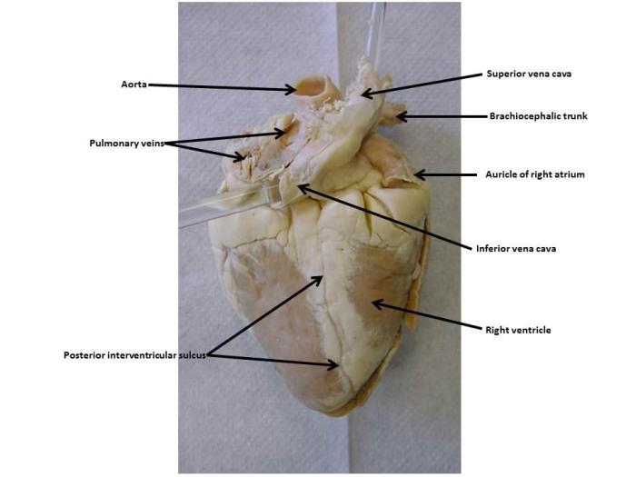
Understanding the anterior surface of the sheep heart is crucial for various clinical applications, particularly in diagnostic and surgical procedures.
In diagnostic procedures, knowledge of the heart’s anatomy is essential for:
Echocardiography
- Echocardiography utilizes ultrasound waves to visualize the heart’s structure and function. Understanding the anterior surface allows accurate positioning of the ultrasound probe to obtain optimal images of the heart chambers, valves, and major vessels.
Cardiac Catheterization
- Cardiac catheterization involves inserting a thin tube (catheter) into the heart’s chambers and vessels. Knowledge of the anterior surface helps guide the catheter safely to its intended location, reducing the risk of complications.
In surgical procedures, understanding the anterior surface of the heart is critical for:
Cardiac Transplantation
- During cardiac transplantation, the diseased heart is replaced with a healthy donor heart. Familiarity with the anterior surface ensures precise identification and connection of the heart’s vessels and structures during the transplantation process.
Valve Replacement Procedures
- Valve replacement procedures involve repairing or replacing damaged heart valves. Understanding the anterior surface provides a clear view of the valves, allowing surgeons to access and operate on them effectively.
Questions and Answers
What are the main structures visible on the anterior view of the sheep heart?
The right and left atria, right and left ventricles, coronary arteries, and pulmonary artery.
What is the function of the pericardial sac surrounding the heart?
To protect and support the heart, preventing excessive movement and providing lubrication.
How is knowledge of the anterior view of the sheep heart used in clinical practice?
It guides diagnostic procedures (echocardiography, cardiac catheterization) and surgical interventions (transplantation, valve replacement).
